45 the brain diagram with labels
How to draw human brain/ draw labelled diagram of brain/brain ... - YouTube Please watch: "cell structure and functions / animal cell vs plant cell / parts of cell / ch 8 science class 8 cbse" ... Parts of the brain: Learn with diagrams and quizzes | Kenhub Labeled brain diagram. First up, have a look at the labeled brain structures on the image below. Try to memorize the name and location of each structure, then proceed to test yourself with the blank brain diagram provided below. Labeled diagram showing the main parts of the brain.
Label the Brain Anatomy Diagram Flashcards | Quizlet Start studying Label the Brain Anatomy Diagram. Learn vocabulary, terms, and more with flashcards, games, and other study tools.

The brain diagram with labels
Picture of the Brain - WebMD The brain is one of the largest and most complex organs in the human body. It is made up of more than 100 billion nerves that communicate in trillions of connections called synapses. • The ... wikieducator.org › Nervous_System_Worksheet_AnswersNervous System Worksheet Answers - WikiEducator Jan 14, 2008 · 8. The diagram below shows a section of a dog’s brain. Add the labels in the list below and, if you like, colour in the diagram as suggested. Cerebellum - blue; Spinal cord - green; Medulla oblongata - orange; Hypothalamus - purple; Pituitary gland - red; Cerebral hemispheres – yellow. 9. Match the descriptions below with the terms in the list. Brain Anatomy and How the Brain Works - Hopkins Medicine The cerebellum ("little brain") is a fist-sized portion of the brain located at the back of the head, below the temporal and occipital lobes and above the brainstem. Like the cerebral cortex, it has two hemispheres. The outer portion contains neurons, and the inner area communicates with the cerebral cortex.
The brain diagram with labels. Structure, Diagram, Parts Of Human Brain - BYJUS The human brain controls nearly every aspect of the human body ranging from physiological functions to cognitive abilities. It functions by receiving and sending signals via neurons to different parts of the body. The human brain, just like most other mammals, has the same basic structure, but it is better developed than any other mammalian brain. Brain Diagram With Labels Pictures, Images and Stock Photos Browse 457 brain diagram with labels stock photos and images available, or start a new search to explore more stock photos and images. Newest results. Brain section 1 Schematic illustration of human cerebrum. Made in vector, easy recolor. brain diagram with labels stock illustrations. Brain section 1. Label Brain Diagram Printout - EnchantedLearning.com The Brain. Read the definitions below, then label the brain anatomy diagram. Cerebellum - the part of the brain below the back of the cerebrum. It regulates balance, posture, movement, and muscle coordination. Corpus Callosum - a large bundle of nerve fibers that connect the left and right cerebral hemispheres. Brain diagram labels Images, Stock Photos & Vectors | Shutterstock Find Brain diagram labels stock images in HD and millions of other royalty-free stock photos, illustrations and vectors in the Shutterstock collection. Thousands of new, high-quality pictures added every day.
Label The Brain - Mr. Barth's Class Label The Brain. The following websites are to help you learn and remember the parts of the brain and their locations. Please go through each of websites and become familiar with each of the parts of the brain. I would advise you to repeat each of them a few times until you have the locations memorized. Click on the link to the left to review ... developingchild.harvard.edu › resources › the-brainThe Brain Circuits Underlying Motivation: An Interactive Graphic The brain systems that govern motivation are built over time, starting in the earliest years of development. These intricate neural circuits and structures are shaped by interactions between the experiences we have and the genes we are born with, which together influence both how our motivation systems develop and how they function later in life. PDF The Human Brain Diagram - Therapist Aid The Human Brain Author: Therapist Aid LLC Created Date: 8/3/2020 5:10:53 PM ... byjus.com › biology › diagram-of-heartHeart Diagram with Labels and Detailed Explanation - BYJUS The diagram of heart is beneficial for Class 10 and 12 and is frequently asked in the examinations. A detailed explanation of the heart along with a well-labelled diagram is given for reference. Well-Labelled Diagram of Heart. The heart is made up of four chambers: The upper two chambers of the heart are called auricles.
Label The Brain Anatomy Diagram [3no75e5ezeld] Label the Brain Anatomy Diagram The Brain Read the definitions below, then label the brain anatomy diagram. Cerebellum - the part of the brain below the back of the cerebrum. It regulates balance, posture, movement, and muscle coordination. Corpus Callosum - a large bundle of nerve fibers that connect the left and right cerebral hemispheres. Nervous System - Label the Brain - TheInspiredInstructor.com This brain part controls thinking. This brain part controls balance, movement, and coordination. This brain part controls involuntary actions such as breathing, heartbeats, and digestion. This part of the nervous system moves messages between the brain and the body. This part of the cerebrum interprets and sorts information from the senses. Brain Label (Remote) - The Biology Corner Brain Label (Remote) Shannan Muskopf December 29, 2020. This brain labeling activity was created for remote learners as an alternative to the labeling and coloring worksheet we would traditionally do in class. Instead of coloring and labeling on printouts, students use google slides to drag labels to the images or type the answers into text boxes. Diagram Of Brain with their Labelings and Detailed Explanation The diagram of the brain is useful for both Class 10 and 12. It is one among the few topics having the highest weightage of marks and is frequently asked in the examinations. A well-labelled diagram of a human brain is given below for further reference. Structure And Function Of The Human Brain.
Spinal Cord Histology Labeled - Top Label Maker
PDF Brain Anatomy - Wou BI 335 - Advanced Human Anatomy and Physiology Western Oregon University Figure 4: Mid-sagittal section of brain showing diencephalon (includes corpus callosum, fornix, and anterior commissure) Marieb & Hoehn (Human Anatomy and Physiology, 9th ed.) - Figure 12.10 Exercise 2: Utilize the model of the human brain to locate the following structures / landmarks for the

my red crayon: put your thinking caps on.
Anatomical diagrams of the brain - e-Anatomy - IMAIOS These original illustrations and diagrams of the brain were created from 3D medical imaging reconstructions and then redrawn and colored using Adobe Illustrator. ... use of interactive anatomical labels. The user can select to display multiple categories of labels on the illustrations: Cerebral lobes / regions; Cerebrum, divided into: frontal ...
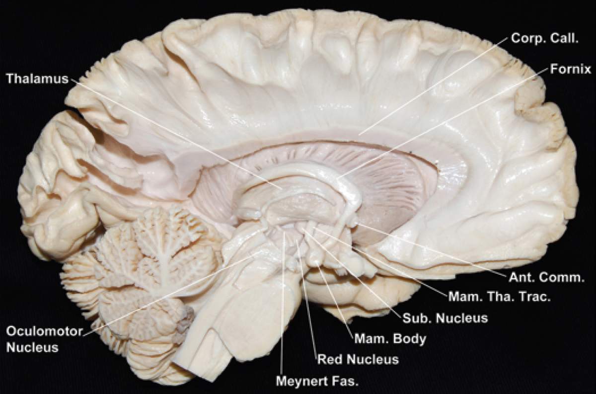
Medial Surface of the Left Hemisphere and Brainstem | Neuroanatomy | The Neurosurgical Atlas, by ...
Diagram of the Brain and its Functions - Bodytomy Given below is a labeled diagram showing the brain stem and its related structures. Brain Stem and Structures. Cerebellum. The word 'cerebellum' literally means little brain. It is the second largest part of the brain, and is located at the back, below the occipital lobe, beneath the cerebrum and behind the brain stem. It contains an outer ...

TooSogiE Medical Images: Cranial Nerves : X - XII
en.wikipedia.org › wiki › File:Human_skeleton_frontFile:Human skeleton front en.svg - Wikipedia Restructured the image internals by adding layers, changing groupings, and adding meaningful ids and labels so that the image is easier to manipulate programmatically. Also made the labels text elements and gave them ids (it might be possible to generate : 10:17, 1 October 2007: 436 × 842 (764 KB) LadyofHats: some changes asked in FP discussion
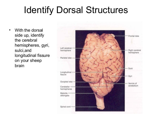
Sheep+brain+dissection
› photos › diagram-of-bodyDiagram Of Body Organs Female Pics Pictures, Images ... - iStock Human internal organs Internal organs in woman and man body. Brain, stomach, heart, kidney, medical icon in female and male silhouette. Digestive, respiratory, cardiovascular systems. Anatomy poster vector illustration. diagram of body organs female pics stock illustrations
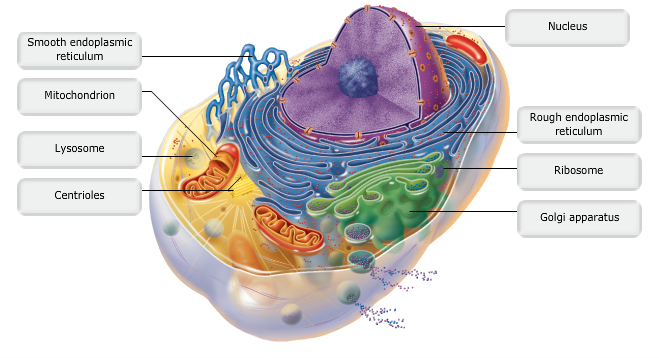
Print A&P Chapter 3 Cells: The Living Units flashcards | Easy Notecards
› enAnatomy, medical imaging and e-learning for ... - IMAIOS IMAIOS and selected third parties, use cookies or similar technologies, in particular for audience measurement. Cookies allow us to analyze and store information such as the characteristics of your device as well as certain personal data (e.g., IP addresses, navigation, usage or geolocation data, unique identifiers).

Midsagittal section of brain Quiz
The Brain Diagram Labeled Pictures, Images and Stock Photos Search from The Brain Diagram Labeled stock photos, pictures and royalty-free images from iStock. Find high-quality stock photos that you won't find anywhere else.

Brain Human Anatomy · Free vector graphic on Pixabay
label the brain anatomy diagram brain sheep labeled worksheet dissection parts lab anatomy diagram label human companion lateral locate diagrams following items help use worksheeto. ... Brain sheep label anatomy labeled superior classroom labels sdmesa physiology nervous inferior edu lateral sup ventricle bottom section sagittal creative. Nervous system worksheet answers.
Post a Comment for "45 the brain diagram with labels"