43 knee joint with labels
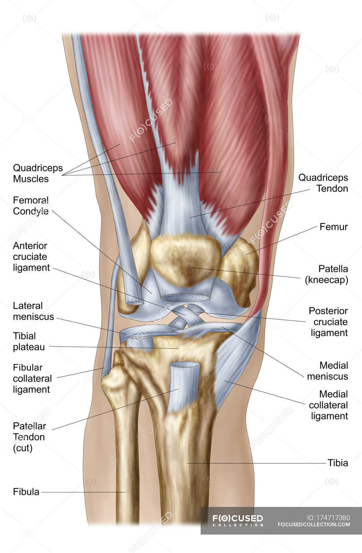
The Knee Joint - Articulations - Movements - Injuries The knee joint is a hinge type synovial joint, which mainly allows for flexion and extension (and a small degree of medial and lateral rotation). It is formed by articulations between the patella, femur and tibia. In this article, we shall examine the anatomy of the knee joint - its articulating surfaces, ligaments and neurovascular supply. Knee Joint - label pictures Flashcards | Quizlet Knee Joint - label pictures Flashcards | Quizlet Knee Joint - label pictures Term 1 / 7 1. Femur 2. Articular capsule 3. PCL 4. Lateral Meniscus 5. ACL 6. Tibia Click the card to flip 👆 Definition 1 / 7 1-6 Click the card to flip 👆 Flashcards Learn Test Match Created by cfreynolds2018 Terms in this set (7) 1. Femur 2. Articular capsule 3. PCL 4.
Amazon.com: OHALEEP Knee Brace, Knee Braces for Knee Pain, … Buy OHALEEP Knee Brace, Knee Braces for Knee Pain, Adjustable Compression Knee Support Braces for ACL, PCL, MCL, Joint Pain Relief Injury Recovery Meniscus Tear,Men and Women on ... We recommend that you do not solely rely on the information presented and that you always read labels, warnings, and directions before using or consuming a ...

Knee joint with labels
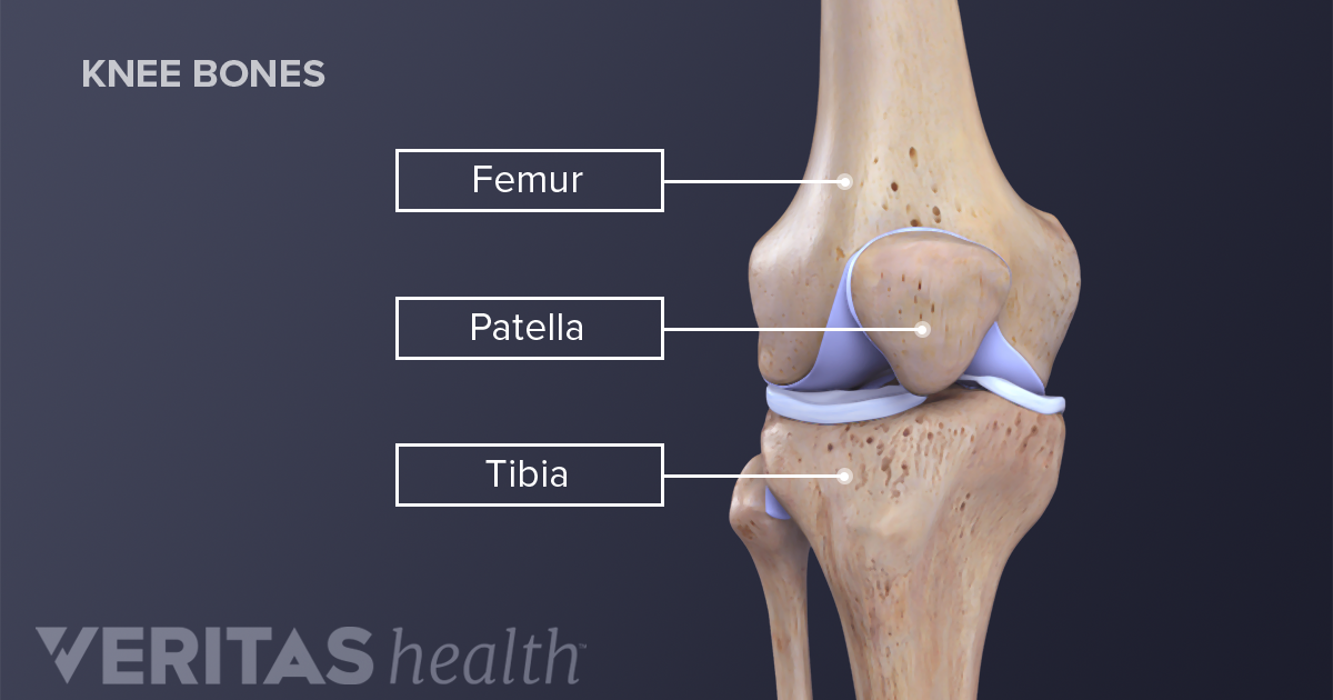
Knee Joint Anatomy: Bones, Ligaments, Muscles, Tendons, Function The knee joint is a synovial joint which connects the femur (thigh bone), the longest bone in the body, to the tibia (shin bone). There are two main joints in the knee: 1) the tibiofemoral joint where the tibia meet the femur 2) the patellofemoral joint where the kneecap (or patella) meets the femur. These two joints work together to form a ... Knee Anatomy, Diagram & Pictures | Body Maps - Healthline The knee is the meeting point of the femur (thigh bone) in the upper leg and the tibia (shinbone) in the lower leg. The fibula (calf bone), the other bone in the lower leg, is connected to the... Anatomy of human knee joint with labels — Stock Photo $449 "Anatomy of human knee joint with labels" is an authentic stock image by StocktrekImages. It's available in the following resolutions: 1049 x 1600px, 1704 x 2600px, 3422 x 5220px. The minimum price for an image is 49$. Image in the highest quality is 3422 x 5220px, 300 dpi, and costs 449$. Similar Images Same Series Keywords
Knee joint with labels. label the knee Quiz - PurposeGames.com This is an online quiz called label the knee There is a printable worksheet available for download here so you can take the quiz with pen and paper. Your Skills & Rank Total Points 0 Get started! Today's Rank -- 0 Today 's Points One of us! Game Points 13 You need to get 100% to score the 13 points available Add to Playlist Sport Medicine Braces, Supports & Recovery Gear | McDavidUSA Shoulder/AC Joint Separation Compression Browse All Compression Arm Sleeves Leg Sleeves Tops Bottoms Socks Outlet ... Knee Brace w/ Polycentric Hinges & Cross Straps $89.99. Knee Support/Double Wrap $37.99. Flex Ice Therapy Ankle Compression Sleeve $34.99. ELITE Bio-Logix™ Ankle Brace $64.99. Knee Joint - San Diego Mesa College Knee Joint. Click on a photo for a larger view of the model. Click on L abel for the labeled model. Back to Muscular System. Anterior: Anterior without patella: Posterior: Label: Label: Label : Label: Label: Knee joint: anatomy, ligaments and movements - Kenhub The tibiofemoral joint is an articulation between the lateral and medial condyles of the distal end of the femur and the tibial plateaus, both of which are covered by a thick layer of hyaline cartilage .
Osteoarthritis knee pain: Foods to eat and avoid Osteoarthritis (OA) of the knee damages the cartilage in the knee joint. Cartilage is a tissue that acts as a cushion at the ends of bones within joints. This results in pain and mobility problems. Amazon.com: OHALEEP Knee Brace, Knee Braces for Knee Pain ... OHALEEP Knee Brace, Knee Braces for Knee Pain, Adjustable Compression Knee Support Braces for ACL, PCL, MCL, Joint Pain Relief Injury Recovery Meniscus Tear,Men and Women Brand: OHALEEP 4.5 out of 5 stars 70 ratings Four-Bar Linkages - University of Illinois Urbana-Champaign Example: Knee joint (constrained motion) The human knee joint is a type of biological hinge , which allows movement in only one primary angle. The knee connects the femur (the upper leg bone) to the tibia (the larger of the two lower leg bones). What are the Parts of the Knee Joint? | Systems4Knees™ There are actually two joints in the knee. The patellofemoral joint is where the femur and the kneecap meet and move together, and the tibiofemoral joint is the point joins the tibia and the femur. These joints help the knees hinge back and forth and move side to side. A diagram showing the bones in and around the knee joint.
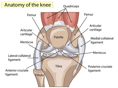
Pain Above Knee: Causes, Treatment, and Prevention - Healthline Apr 01, 2019 · Arthritis in your knee occurs when the cartilage supporting your knee joint wears away. Common types of arthritis such as osteoarthritis , rheumatoid arthritis , and lupus can all cause pain ... Labeling the Knee Joint Quiz - PurposeGames.com This is an online quiz called Labeling the Knee Joint There is a printable worksheet available for download here so you can take the quiz with pen and paper. Your Skills & Rank Total Points 0 Get started! Today's Rank -- 0 Today 's Points One of us! Game Points 11 You need to get 100% to score the 11 points available Actions Add to favorites A Diagrammatic Explanation of the Parts of the Human Knee Knee actually consists of three bones - femur, tibia and patella. Femur is the thigh bone, tibia is the shin bone and patella is the small cap like structure which rests on the other two bones. Femur is considered as the largest bone in the human body. The femur and the tibia meets at the tibiofemoral joint and patella rests on top of this joint. Knee Anatomy: Bones, Muscles, Tendons, and Ligaments - Verywell Health Bones Around the Knee There are three important bones that come together at the knee joint: The tibia (shin bone) The femur (thigh bone) The patella (kneecap) A fourth bone, the fibula, is located just next to the tibia and knee joint, and can play an important role in some knee conditions.
label the knee Quiz
NIfTI: — Neuroimaging Informatics Technology Initiative Dec 18, 2013 · The primary goal of NIfTI is to provide coordinated and targeted service, training, and research to speed the development and enhance the utility of informatics tools related to neuroimaging. The National Institute of Mental Health and the National Institute of Neurological Disorders and Stroke are joint sponsors of this initiative.

Knee x-ray - labeling questions | Radiology Case ...
A Labeled Diagram of the Knee With an Insight into Its Working Labeled Diagram of the Knee Joint Knee joint is one of the most important hinge joints of our body. Its complexity and its efficiency is the best example of God's creation. The anatomy of the knee consists of bones, muscles, nerves, cartilages, tendons and ligaments. All these parts combine and work together.

Label the parts of the knee joint. | Homework.Study.com
Pain Above Knee: Causes, Treatment, and Prevention - Healthline 01.04.2019 · Pain above your knee may indicate a number of potential conditions. Learn about what causes this pain, how it's treated, and when to seek medical attention.
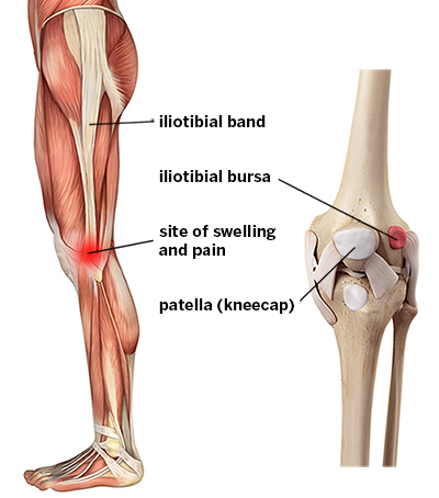
IT Band Syndrome: Knee Pain Symptoms & Treatments | HSS
Labeling The Knee Joint Quick and Easy Solution Let me give you a short tutorial. Read! Don't miss. Step 1. Go to Labeling The Knee Joint website using the links below Step 2. Enter your Username and Password and click on Log In Step 3. If there are any problems, here are some of our suggestions Top Results For Labeling The Knee Joint Updated 1 hour ago

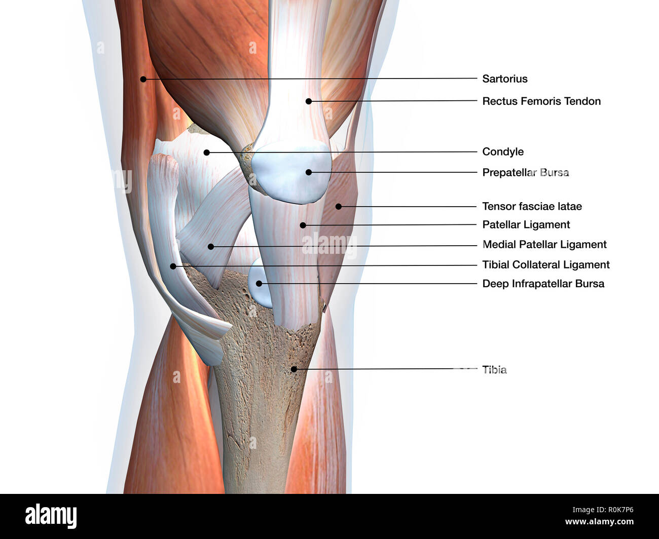
Knee Muscles And Ligaments Parts Labeled On White Background ...
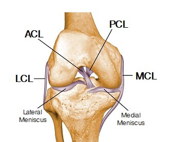
Knee Ligaments: Anatomy, ACL, MCL, PCL, LCL, Torn Ligament An injury to a knee ligament is called a sprain or a tear. Many knee sprains are mild, but torn knee ligaments can be severe. Knee ligament injuries are common, especially in athletes. The ligaments can be overstretched or torn when: Force is applied to the back of the knee when the joint is partly flexed.
Synovial joint - Teaching resources
Knee Images and Pictures - Photos and X-Rays of the Knee - Verywell Health The knee is one of the most commonly injured joints in the body. The knee joint is the junction of the thigh and the leg (part of the lower extremity). The femur (thigh bone) contacts the tibia (shin bone) at the knee joint. The patella (kneecap) sits over the front of the knee joint. Four major ligaments connect the bones and stabilize the ...
Knee Anatomy Images – Browse 33,429 Stock Photos, Vectors ...
Knee Joint Picture Image on MedicineNet.com The thigh bone (the femur) meets the large shin bone (the tibia) to form the main knee joint. This joint has an inner (medial) and an outer (lateral) compartment. The kneecap (the patella) joins the femur to form a third joint, called the patellofemoral joint. The patella protects the front of the knee joint.
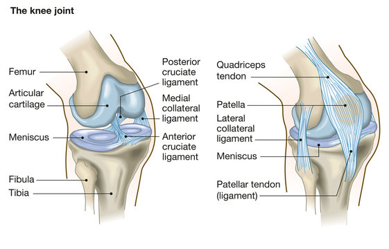
Knee Joint
Alila Medical Media | Knee joint, basic labels Human knee joint diagram showing joint cavity, capsule, all cartilage. - Alila Medical Media.
Knee joint: anatomy, ligaments and movements | Kenhub
Radiopaedia - Drawing Bones of the knee joint - English labels Description: Bones of the knee joint. This drawing shows the different parts of the bones of the knee. English labels. Case courtesy of Dr Henry Knipe, Radiopaedia.org. From the case rID: 31397. Anatomical structures in item: Genu. Articulatio genus.
Patella Bone - Anterior and Posterior Views | GetBodySmart
Knee Joint Labeled Diagram - Dreamstime.com Knee Joint Labeled Diagram Royalty-Free Stock Photo A knee joint with detailed labels anatomy knee, knee anatomy, joint cartilage, detailed labels, knee, labels, joint, doctor, health, anatomy, medicine, pain, osteoporosis, arthritis, disease, bone, leg, tear, femur, cartilage, disc, shading, acl, ligament, tibia More ID 39627491
Joints
Amazon.com: Hempactiv Joint & Muscle Relief Cream, Infused ... Hempactiv Joint & Muscle Relief Cream, Infused with Hemp, Menthol, MSM & Arnica, Soothe Discomfort in Your Back, Muscles, Joints, Neck, Shoulder, Knee, Nerves - 2 Fl Oz Brand: HEMPACTIV 4.5 out of 5 stars 10,862 ratings
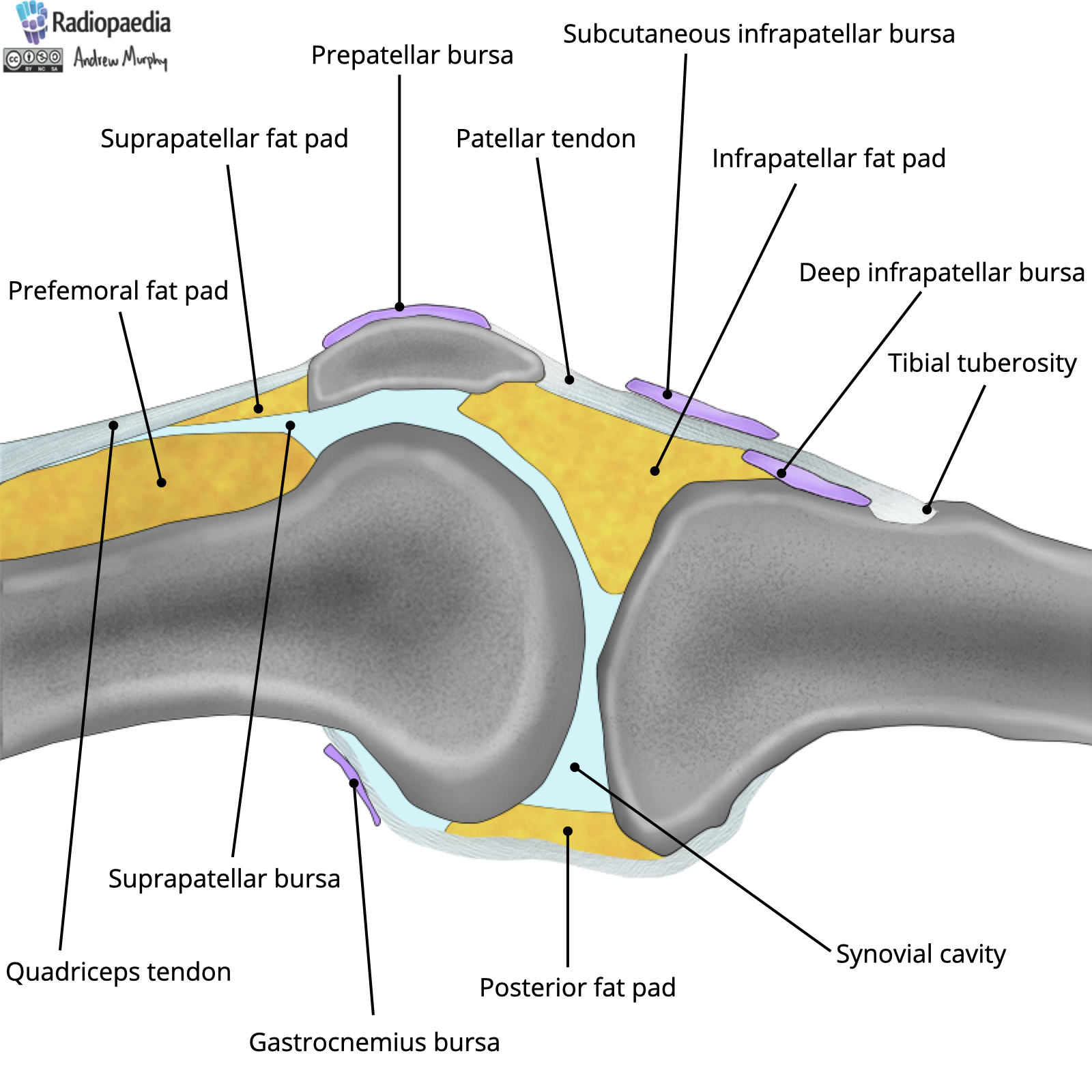
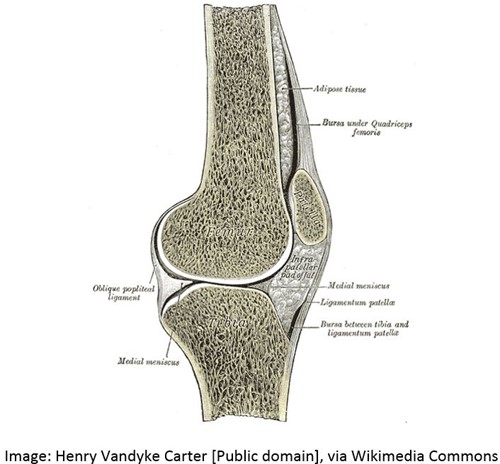
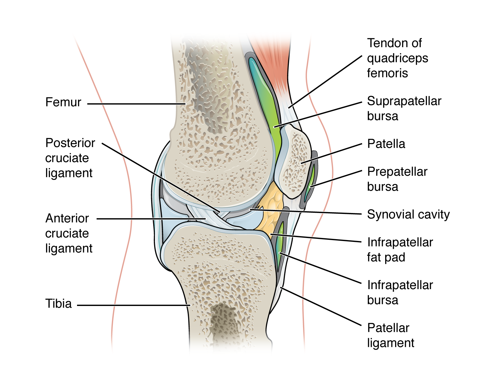
Radiopaedia - Drawing Fat pads and bursae of the knee ...
3D Knee Joint Model *Finished Product* - YouTube Finally completed my knee joint model with labels of all the key ligaments, muscles, tendons, and bursae. Let me know what you think, I spent a lot of time ...
Knee | Clinical Gate
Sport Medicine Braces, Supports & Recovery Gear | McDavidUSA Flex Ice Therapy Knee/Thigh Compression Sleeve $39.99 Phantom Ankle Brace w/ Advanced Strapping & Flex-Support Stirrup Stays $39.99 Knee Brace w/ Polycentric Hinges & Cross Straps $89.99

9.4 Synovial Joints – Anatomy & Physiology
Solved Label the structures of the knee joint (anterior - Chegg Expert Answer. 88% (8 ratings) Transcribed image text: Label the structures of the knee joint (anterior view) by clicking and dragging the labels to the correct location. Fibula Tibial collateral ligament Tibial tuberosity Medial meniscus Posterior cruciate ligament Lateral condyle with articular cartilage Lateral meniscus Femur Medial condyle ...
Chondromalacia Patellae - Children's Health Issues - Merck ...
NIfTI: — Neuroimaging Informatics Technology Initiative 18.12.2013 · The primary goal of NIfTI is to provide coordinated and targeted service, training, and research to speed the development and enhance the utility of informatics tools related to neuroimaging. The National Institute of Mental Health and the National Institute of Neurological Disorders and Stroke are joint sponsors of this initiative.

Solved Click and drag the labels to their correct locations ...
Amazon.com: anatomical model knee LYOU Human Knee Joint Model with Knee Ligament, Life Size Anatomical Knee Joint Flexible Skeleton Model, Perfect for Medical Learning and Teaching 9 $3998 Get it as soon as Thu, Apr 14 FREE Shipping by Amazon Wellden Product Anatomical Human Knee Joint Model, w/Ligaments, Functional, Life Size 9 $4500 Get it Tue, Apr 19 - Mon, Apr 25
Knee joint: anatomy, ligaments and movements | Kenhub
Knee Joint Anatomy: Structure, Function & Injuries The specific design of knee joint anatomy allows a number of functions: Supports the body in upright position without muscles having to work. Helps in lowering and raising body e.g. sitting, climbing and squatting. Allows rotation/twisting of the leg to place and position foot accurately.
Knee Joint Anatomy: Structure, Function & Injuries - Knee ...
Knee x-ray - labeling questions | Radiology Case - Radiopaedia Normal X-ray Knee - Frontal (with labels) Annotated image Annotated image Frontal Knee Frontal 1. Femoral shaft 2. Patella 3. Base of patella 4. Apex of patella 5. Adductor tubercle of femur 6. Medial epicondyle of femur 7. Medial condyle of femur 8. Lateral epicondyle of femur 9. Lateral condyle of femur 10. Groove for popliteus 11.
Knee Anatomy
Osteoarthritis knee pain: Foods to eat and avoid - Medical News … Learning how to read the nutritional labels can help a person to avoid high levels of salt, sugar, and unhealthful fats. Last medically reviewed on October 9, 2018 Osteoarthritis

Diagrams - Knee Joint Quiz - By mattandersen91
Knee Joint Label Flashcards | Quizlet Knee Joint Label Created by LaLaKub91 Terms in this set (10) femur What is A? lateral collateral ligament what is d? lateral meniscus what is e? fibula what is g? tibia what is h? posterior cruciate ligament What is j? anterior cruciate ligament what is k? medial meniscus what is l? medial collateral ligament what is m? Patella what is n?

Knee x-ray - labeling questions | Radiology Case ...
Amazon.com: Hempactiv Joint & Muscle Relief Cream, Infused … Buy Hempactiv Joint & Muscle Relief Cream, Infused with Hemp, Menthol, MSM & Arnica, Soothe Discomfort in Your Back, Muscles, Joints ... We recommend that you do not solely rely on the information presented and that you always read labels, warnings, and directions before using or consuming a product. For additional information about a ...

Knee - Wikipedia
Knee Joint Anatomy Labeled Stock Illustration 157672166 Find Knee Joint Anatomy Labeled stock images in HD and millions of other royalty-free stock photos, illustrations and vectors in the Shutterstock collection ...
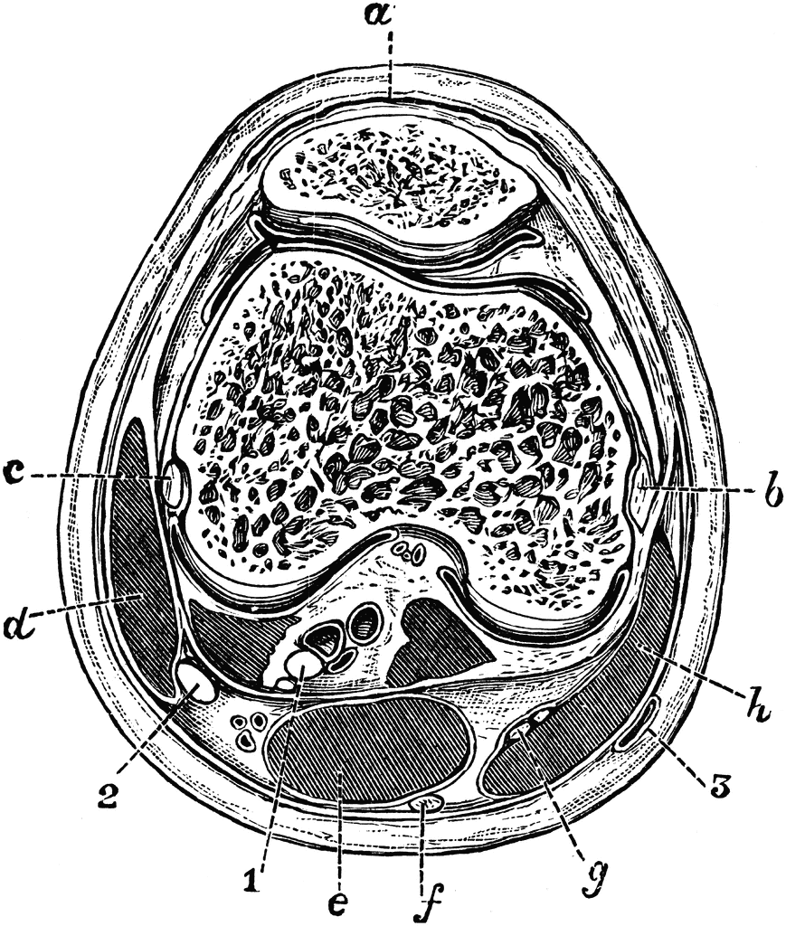
Transverse Section of the Knee Joint | ClipArt ETC
Knee Anatomical Models | Knee Joint Models - Universal Medical Inc Functional Model of the Knee Joint $314.00 Deluxe Functional Knee Joint Model MSRP $161.00 $148.00 Functional Knee Joint Model MSRP $110.00 $101.00 Rating: 1 Review Ultraflx Ligamented Knee - Functional Replica $204.00 Knee Joint with Ligaments Model $108.00 Knee Joint with Removable Muscles 12-Part MSRP $510.00 $469.00

The knee – everything about the anatomy of the knee joint | medi
Knee Joint - Anatomy Pictures and Information - Innerbody The knee, also known as the tibiofemoral joint, is a synovial hinge joint formed between three bones: the femur, tibia, and patella. Two rounded, convex processes (known as condyles) on the distal end of the femur meet two rounded, concave condyles at the proximal end of the tibia. Continue Scrolling To Read More Below... Additional Resources

File:917 Knee Joint.jpg - Wikimedia Commons
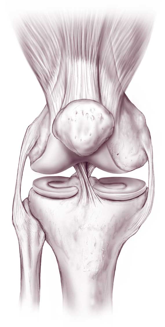
Radiopaedia - Drawing Ligaments of the knee joint - no labels None available. Description: Ligaments of the knee joint. This drawing shows the different ligaments of the knee. Case courtesy of Dr Henry Knipe, Radiopaedia.org. From the case rID: 31397. Anatomical structures in item:
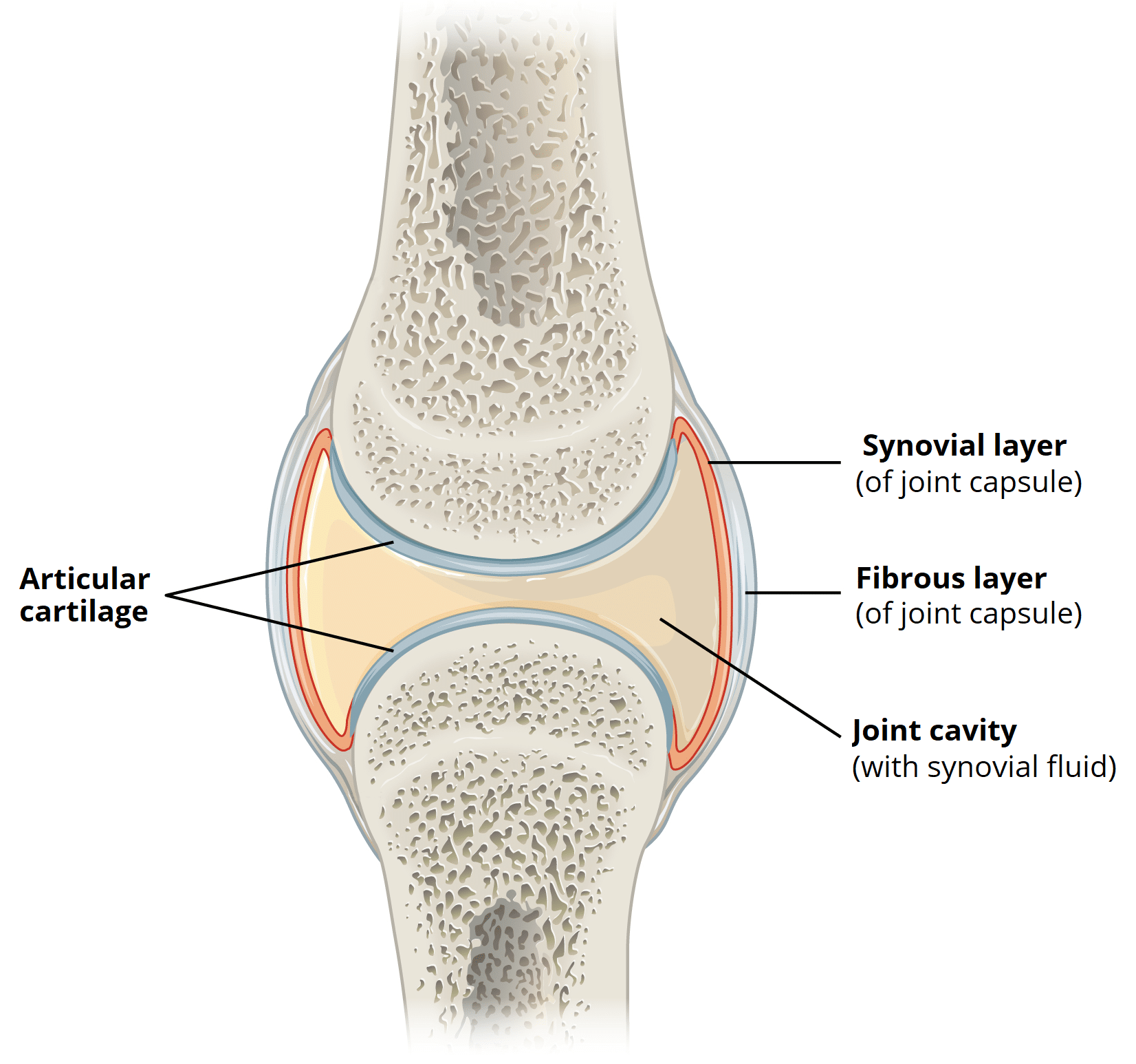
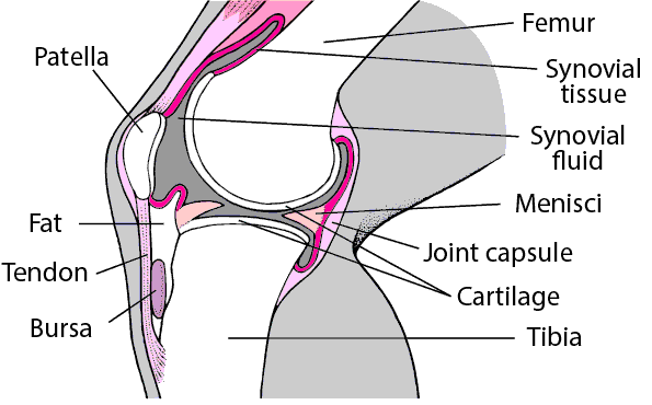
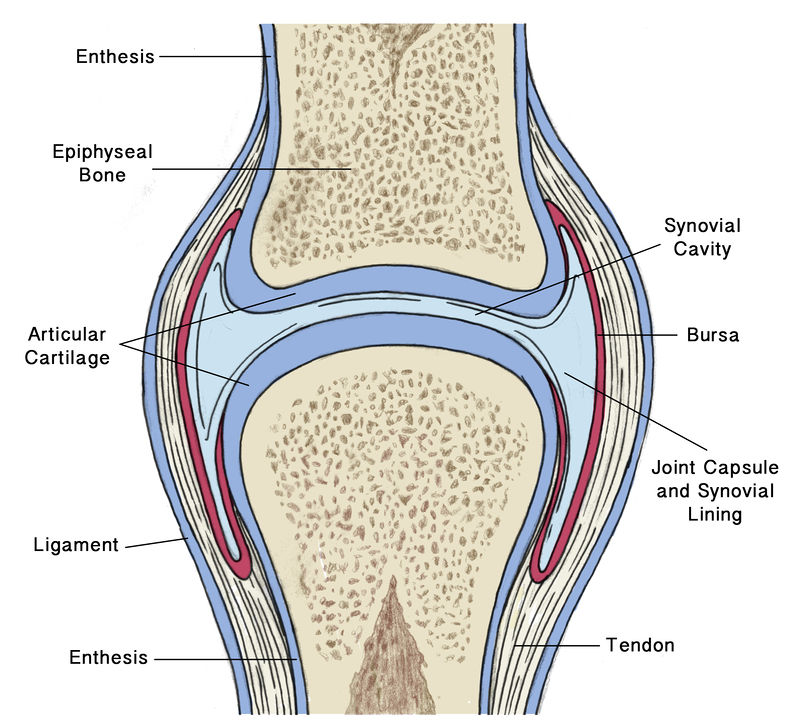
Structures of a Synovial Joint - Capsule - Ligaments ...
Four-Bar Linkages - University of Illinois Urbana-Champaign Example: Knee joint (constrained motion) The human knee joint is a type of biological hinge, which allows movement in only one primary angle. The knee connects the femur (the upper leg bone) to the tibia (the larger of the two lower leg bones). These two bones sit next to each other and are free to rotate about a single axis.

Labeling the Knee Joint Quiz
Anatomy of human knee joint with labels — Stock Photo $449 "Anatomy of human knee joint with labels" is an authentic stock image by StocktrekImages. It's available in the following resolutions: 1049 x 1600px, 1704 x 2600px, 3422 x 5220px. The minimum price for an image is 49$. Image in the highest quality is 3422 x 5220px, 300 dpi, and costs 449$. Similar Images Same Series Keywords

The Knee Joint Laminated Anatomy Chart
Knee Anatomy, Diagram & Pictures | Body Maps - Healthline The knee is the meeting point of the femur (thigh bone) in the upper leg and the tibia (shinbone) in the lower leg. The fibula (calf bone), the other bone in the lower leg, is connected to the...
Anatomy of human knee joint with labels — text, bones - Stock ...
Knee Joint Anatomy: Bones, Ligaments, Muscles, Tendons, Function The knee joint is a synovial joint which connects the femur (thigh bone), the longest bone in the body, to the tibia (shin bone). There are two main joints in the knee: 1) the tibiofemoral joint where the tibia meet the femur 2) the patellofemoral joint where the kneecap (or patella) meets the femur. These two joints work together to form a ...
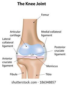
Knee Joint Labeled Diagram Stock Illustration 186348857 ...
Knee joint showing muscles and ligaments with labels Stock ...
Knee Anatomy Images – Browse 33,429 Stock Photos, Vectors ...
Leg and knee anatomy: Bones, muscles, soft tissues | Kenhub

Left: The initial label configuration for the knee joint. The ...
Introducing the knee: Anatomy and biomechanics ...
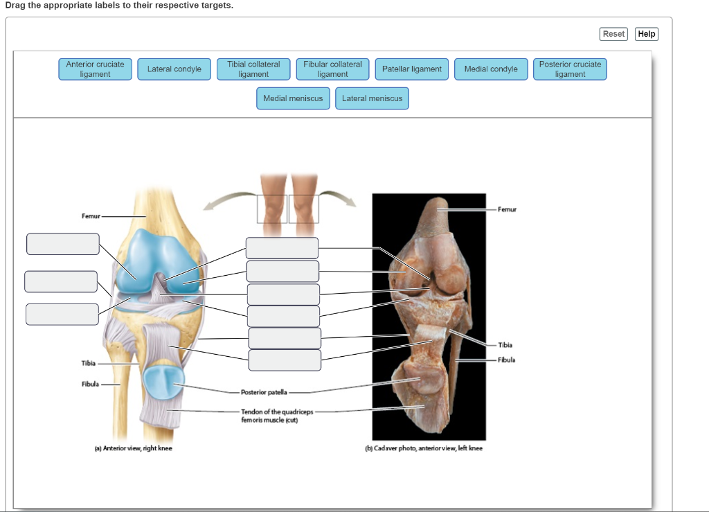
Solved Drag the appropriate labels to their respective ...

Broken Knee Fracture stock vector. Illustration of articular ...

Knee joint | Alila Medical Images

Knee Joint Picture Image on MedicineNet.com

knee joint diagram labelled - Clip Art Library
Human Biology fig. 1.15 - Synovial joint - English labels ...
9.4 Synovial Joints – Anatomy & Physiology
Knee Joint Anatomy: Bones, Ligaments, Muscles, Tendons, Function
Post a Comment for "43 knee joint with labels"